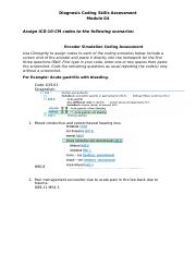
What is the ICD 9 code for exudative macular Degen?
Short description: Exudative macular degen. ICD-9-CM 362.52 is a billable medical code that can be used to indicate a diagnosis on a reimbursement claim, however, 362.52 should only be used for claims with a date of service on or before September 30, 2015.
Which ICD 10 code should not be used for reimbursement purposes?
H35.32 should not be used for reimbursement purposes as there are multiple codes below it that contain a greater level of detail. The 2022 edition of ICD-10-CM H35.32 became effective on October 1, 2021.
What is the new ICD 10 for nexdtve?
Short description: Nexdtve age-related mclr degn, bilateral, early dry stage The 2022 edition of ICD-10-CM H35.3131 became effective on October 1, 2021. This is the American ICD-10-CM version of H35.3131 - other international versions of ICD-10 H35.3131 may differ.
What is the difference between ICD-9-CM and CPT?
ICD-9-CM 310.9 is one of thousands of ICD-9-CM codes used in healthcare. Although ICD-9-CM and CPT codes are largely numeric, they differ in that CPT codes describe medical procedures and services. Can't find a code?

What is ICD 9 code for macular degeneration?
362.52Macular degeneration generally starts in the dry form and is classified to ICD-9-CM code 362.51. Wet macular degeneration (362.52) is characterized by the growth of abnormal blood vessels from the choroid underneath the macula.
What is the ICD-10 for macular degeneration?
Unspecified macular degeneration H35. 30 is a billable/specific ICD-10-CM code that can be used to indicate a diagnosis for reimbursement purposes. The 2022 edition of ICD-10-CM H35. 30 became effective on October 1, 2021.
How is macular degeneration diagnosed?
Age-related macular degeneration can be detected in a routine eye exam, which will include having your eyes dialated. One of the most common early signs of macular degeneration is the presence of drusen -- tiny yellow deposits under the retina -- or pigment clumping. Your doctor can see these as they examine your eyes.
What is the ICD-10 code for wet AMD?
Table 2: Wet Age-Related Macular Degeneration (AMD)Right EyeLeft EyeWet (exudative) AMD, with active choroidal neovascularizationH35.3211H35.3221Wet (exudative) AMD, with inactive choroidal neovascularizationH35.3212H35.3222Wet (exudative) AMD, inactive scarH35.3213H35.32231 more row
What is exudative macular degeneration?
Exudative macular degeneration is a progressive eye disease that affects the macula or central part of the retina. It causes the eye to develop leaky blood vessels behind the macula, the part of the eye that enables us to see what is straight in front of us.
What is the ICD-10 code for DM with macular degeneration?
E11. 311 - Type 2 diabetes mellitus with unspecified diabetic retinopathy with macular edema | ICD-10-CM.
What are the different types of macular degeneration?
There are two main types of age-related macular degeneration: dry (atrophic) and wet (neovascular or exudative.) In Dry Macular Degeneration, fatty deposits called DRUSEN develop on the macula. Researchers believe that these spots are deposits or debris from deteriorating tissue.
Is there another name for macular degeneration?
Because the disease happens as you get older, it's often called age-related macular degeneration. It usually doesn't cause blindness but might cause severe vision problems. Another form of macular degeneration, called Stargardt disease or juvenile macular degeneration, affects children and young adults.
What are the three most common ways to diagnose macular degeneration?
To confirm a diagnosis of macular degeneration, he or she may do several other tests, including:Examination of the back of your eye. ... Test for defects in the center of your vision. ... Fluorescein angiography. ... Indocyanine green angiography. ... Optical coherence tomography. ... Optical coherence tomography (OCT) angiography.
What is the medical term AMD?
Age-related macular degeneration (AMD) is an eye disease that can blur your central vision. It happens when aging causes damage to the macula — the part of the eye that controls sharp, straight-ahead vision. The macula is part of the retina (the light-sensitive tissue at the back of the eye).
What does Armd stand for in medical terms?
Also called age-related macular degeneration, AMD, and macular degeneration.
What is neovascular age-related macular degeneration?
Neovascular AMD is an advanced form of macular degeneration that historically has accounted for the majority of vision loss related to AMD. The presence of choroidal neovascular membrane (CNV) formation is the hallmark feature of neovascular AMD.
What are early warning signs of macular degeneration?
SymptomsVisual distortions, such as straight lines seeming bent.Reduced central vision in one or both eyes.The need for brighter light when reading or doing close-up work.Increased difficulty adapting to low light levels, such as when entering a dimly lit restaurant.Increased blurriness of printed words.More items...•
At what age does macular degeneration usually begin?
Age-related macular degeneration usually begins at age 55 or older. There is a very low risk of progression from the early stage to the late stage of AMD (which involves vision loss) within five years after diagnosis.
Can Opticians diagnose macular degeneration?
The optometrist at your local optician's practice can test sight, prescribe glasses and check for eye disease. Some optometrists use photography or other imaging to detect early signs of macular degeneration. These might include optical coherence tomography (OCT) scans which create cross-sectional images of the retina.
How long does it take to go blind with macular degeneration?
In late stages of AMD, you may have difficulty seeing clearly. On average, it takes about 10 years to move from diagnosis to legal blindness, but there are some forms of macular degeneration that can cause sight loss in just days. So, please contact us right away if you begin to experience symptoms.
What is the code for AMD wet?
The codes for wet AMD—H35.32xx—use the sixth character to indicate laterality and the seventh character to indicate staging as follows:
Why use a diagnosis code in the absence of an approved therapy?
Why use a diagnosis code in the absence of an approved therapy? Accurate documentation and coding will help researchers and policymakers track the visual impairment and visual function deficits that are associated with the condition. Furthermore, when treatments do become available, you will be ready to code for them.
What is H35.31x3?
H35.31x3 for advanced atrophic dry AMD without subfoveal involvement —geographic atrophy (GA) not involving the center of the fovea.
What is the code for fovea?
The Academy recommends that when coding, you indicate whether the GA involves the center of the fovea: Code H35.31x4 if it does and H35.31x3 if it doesn’t, with “x” indicating lateral ity. Improved categorization of GA will help in clinical practice and also will lead to a better understanding of the natural history, comorbidities, and visual prognosis associated with the disease.
Can an inactive scar be a CNV?
Similarly, an eye that has an inactive scar could have active CNV after the diagnosis of an inactive scar, and treatment can be considered at the time of active CNV. 1 American Academy of Ophthalmology Retina/Vitreous Panel. Preferred Practice Pattern Guidelines: Age-Related Macular Degeneration.

Popular Posts:
- 1. what is the icd 10 code for ppd plant
- 2. icd 10 code for hemarthrosis left knee
- 3. icd-10 code for chest pressure
- 4. icd 10 dx code for fibromuscular dysplasia of renal artery
- 5. icd 10 code for possibly swallowed battery
- 6. icd 10 code for hematuria gross
- 7. icd 10 code for digoxin use
- 8. what is the icd 9 code for acute tonsillitis
- 9. icd 9 code for anx
- 10. icd-10 code for micrognathia