611.72
What is the ICD 9 code for lump or mass in breast?
Lump or mass in breast. ICD-9-CM 611.72 is a billable medical code that can be used to indicate a diagnosis on a reimbursement claim, however, 611.72 should only be used for claims with a date of service on or before September 30, 2015.
What is the ICD-9-CM code for breast cancer?
When ICD-9-CM codes 174.0-174.9, 175.0, or 175.9 are used to identify breast cancer, the patient records must document that the tumor is Clinical Stage I or II.
How are palable breast masses diagnosed in breast cancer?
Palable breast masses are common and usually benign, but efficient evaluation and prompt diagnosis are necessary to rule out malignancy. A thorough clinical breast examination, imaging, and tissue sampling are needed for a definitive diagnosis.
What is the ICD 10 code for mass and lump?
ICD-10-CM Diagnosis Code R22.2. Localized swelling, mass and lump, trunk. 2016 2017 2018 2019 2020 2021 Billable/Specific Code. Type 1 Excludes. intra-abdominal or pelvic mass and lump ( R19.0-) intra-abdominal or pelvic swelling ( R19.0-) Type 2 Excludes. breast mass and lump ( N63) ICD-10-CM Codes Adjacent To N63.
What is the code for breast mass?
"N63. 0 - Unspecified Lump in Unspecified Breast." ICD-10-CM, 10th ed., Centers for Medicare and Medicaid Services and the National Center for Health Statistics, 2018.
What is N63 20 code?
ICD-10 code N63. 20 for Unspecified lump in the left breast, unspecified quadrant is a medical classification as listed by WHO under the range - Diseases of the genitourinary system .
What is diagnosis code N63 11?
ICD-10-CM Code for Unspecified lump in the right breast, upper outer quadrant N63. 11.
What is the CPT code for left breast mass?
Unspecified lump in the left breast, unspecified quadrant N63. 20 is a billable/specific ICD-10-CM code that can be used to indicate a diagnosis for reimbursement purposes. The 2022 edition of ICD-10-CM N63. 20 became effective on October 1, 2021.
What is the ICD-10 code for right breast mass?
ICD-10 code N63. 1 for Unspecified lump in the right breast is a medical classification as listed by WHO under the range - Diseases of the genitourinary system .
What does code Z12 31 mean?
For example, Z12. 31 (Encounter for screening mammogram for malignant neoplasm of breast) is the correct code to use when you are ordering a routine mammogram for a patient. However, coders are coming across many routine mammogram orders that use Z12.
What is diagnosis code N64 4?
ICD-10 code N64. 4 for Mastodynia is a medical classification as listed by WHO under the range - Diseases of the genitourinary system .
What is the ICD 10 code for breast?
Breast Cancer ICD-10 Code Reference SheetLeftC50.012Malignant neoplasm of nipple and areola, left female breastC50.112Malignant neoplasm of central portion, left female breastC50.212Malignant neoplasm of upper-inner quadrant, left female breastC50.312Malignant neoplasm of lower-inner quadrant, left female breast8 more rows
What quadrant is 9 o'clock in the breast?
When looking at the left breast, the upper outer quadrant is between 12 and 3 o'clock; the lower outer quadrant is between 3 and 6 o'clock; the lower inner quadrant is between 6 and 9 o'clock; and the upper inner quadrant is between 9 and 12 o'clock.
What is the ICD-10 code for Mass in left breast?
N63.2ICD-10 Code for Unspecified lump in the left breast- N63. 2- Codify by AAPC.
What is a mass in your breast?
A breast lump is a mass that develops in your breast. While a breast lump can be a sign of breast cancer, often it is not related to cancer. Eight out of 10 breast lumps are noncancerous. If you feel a lump in your breast or under your arm, see your healthcare provider.
What is the ICD-10 for breast ultrasound?
8 for Other abnormal and inconclusive findings on diagnostic imaging of breast is a medical classification as listed by WHO under the range - Symptoms, signs and abnormal clinical and laboratory findings, not elsewhere classified .
What does other abnormal and inconclusive findings on diagnostic imaging of breast mean?
Abnormal mammogram results occur when breast imaging detects an irregular area of the breast that has the potential to be malignant. This could come in the form of small white spots called calcifications, lumps or tumors called masses, and other suspicious areas.
What is the ICD 10 code for abnormal mammogram?
793.80 - Abnormal mammogram, unspecified | ICD-10-CM.
When will the ICd 10 for lumps in breast be effective?
The 2021 edition of ICD-10-CM N63 became effective on October 1, 2020.
When will the ICd 10-CM N63 be released?
The 2022 edition of ICD-10-CM N63 became effective on October 1, 2021.
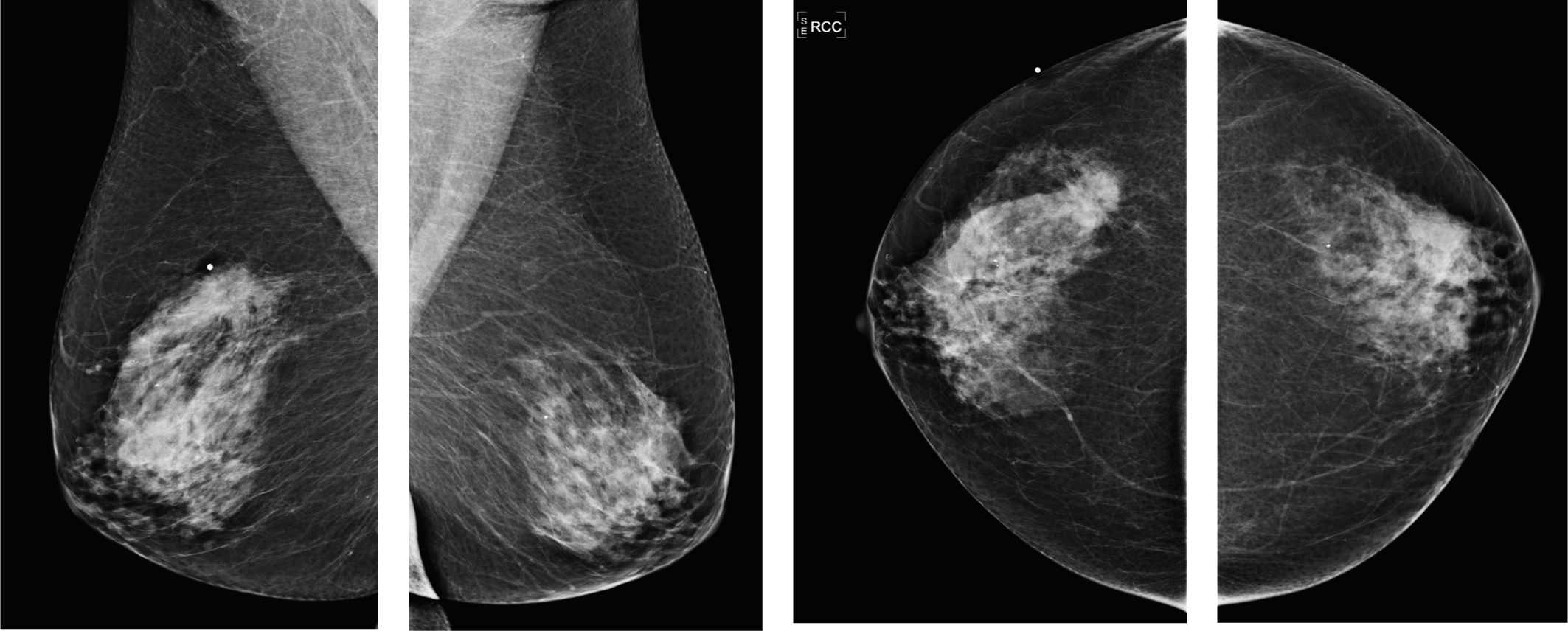
Can a mammary mass be detected?
A mass in the mammary gland, either mobile or immobile. Once the mass has reached the size of a small garden pea, it can be detected by palpation . With mammography a larger number of early breast cancers are being detected since this techniques allows detection prior to the point at which the mass can be felt. Breast masses are not always malignant. Benign fibrocystic breast disease is not uncommon. A fine needle biopsy aspiration can distinguish a cystic mass from a solid one.
What is 19086 breast biopsy?
19086 Biopsy, breast, with placement of breast localization device (s) (e.g., clip, metallic pellet), when performed, and imaging of the biopsy specimen, when performed, percutaneous ; each additional lesion, including magnetic resonance guidance (List separately in addition to code for primary procedure)
How much does a breast biopsy cost?
19083 Biopsy, breast, with placement of breast localization device (s) (e.g., clip, metallic pellet), when performed, and imaging of the biopsy specimen, when performed, percutaneous; first lesion, including ultrasound guidance – average fee payment – $700 – $720
What is SNLB in cancer?
Coding Sentinel Node Biopsy (SNLB) is a surgical procedure in Melanoma and Breast Cancer Screening to determine if cancer has spread beyond a primary tumor into the lymphatic system. Sentinel Node Biopsy in Breast Cancer Evaluation reveals cancer spread, then the patient needs additional lymph nodes removed.
What is the procedure code for breast biopsy?
procedure code and description#N#19081 Biopsy, breast, with placement of breast localization device (s) (e.g., clip, metallic pellet), when performed, and imaging of the biopsy specimen, when performed, percutaneous; first lesion, including stereotactic guidance
Is a sentinel node biopsy more invasive than lymphadenectomy?
You should consider sentinel node biopsy (38500-38530) to be a more “targeted” and less invasive procedure than lymphadenectomy (38700-38780). The sentinel node is the first lymph node to receive drainage from a cancer-containing area of the breast (or other sites).
What is the code for breast ultrasound?
If performing a diagnostic breast ultrasound evaluation and an ultrasound guided needle procedure during the same patient encounter both codes may be billed: the diagnostic ultrasound (76645) and the ultrasound guided biopsy.
Is lymphadenectomy unplanned?
The lymphadenectomy is unplanned at the time of the biopsy. The decision to perform lymphadenectomy (at the same or a later session) is based on the results of the biopsy. Example: The surgeon takes a biopsy of the sentinel axillary node (38525, Biopsy or excision of lymph node [s]; open, deep axillary node [s]).
What is a breast mass?
Breast masses are common and are associated with a wide spectrum of disease, from benign physiological adenosis to advanced malignancy. A structured and comprehensive approach is required in order to assess patients presenting with a new breast mass. This activity outlines the evaluation and management of a new breast mass and highlights the role of the interprofessional team in caring for patients with this condition.
What is the breast imaging report?
Imaging reports are standardized using a tool called BIRADS – Breast Imaging Reporting and Data System (fifth edition). This standard allows breast imaging to be described according to a certain structure: density of breast tissue, presence and location of a mass or masses, calcifications, asymmetry, and any associated features. [23] This classification system divides patients into categories 0 to 6, depending on the likelihood of malignancy in the obtained images:
Why is ultrasound preferred over mammography?
Ultrasound imaging is preferred to mammography in younger women and men as their breast tissue tends to be denser, with a much lower proportion of fatty tissue. This dense tissue impedes the accuracy of mammography and makes it more challenging to detect microcalcifications. [20]
How common is breast cancer?
According to the WHO, breast cancer is the leading cause of cancer-related deaths worldwide, with an estimated lifetime risk of 12%.[3] Benign breast disease is often more common, affecting between 25% and 50% of adult women and accounting for 3% of general practitioners' encounters with female patients.[9] A majority of these cases may present initially with a new breast mass. It is crucial, therefore, for every clinician to have confidence in assessing and managing these patients, and a thorough, consistent approach will enable this. The triple assessment approach discussed in this article has improved outcomes by allowing timely diagnosis and a coordinated interprofessional approach. [10]
What blood test is used for breast abscess?
Baseline blood tests are usually recommended in a patient likely to undergo surgery, emphasizing hemoglobin, bone profile, and liver function tests in case of suspected hepatic metastasis. Inflammatory markers and blood cultures should be considered where a breast abscess is suspected. Tumor markers such as Ca27.29 and Ca15-3 can be used for prognostication and monitoring for recurrence.
What are the most common radiological tools for imaging breast tissue?
The most common radiological tools for imaging breast tissue are mammography, ultrasound, and occasionally MRI.
What is the importance of a thorough history of breast mass?
A thorough and accurate history is the cornerstone of approaching any new breast mass.[12] Particular emphasis should be placed on the chronological development of the lump and the symptoms associated with it.
Popular Posts:
- 1. icd 10 code for small cell carcinoma of right lung
- 2. icd 10 code for activity unarmed fight
- 3. icd 10 code for gave
- 4. icd 10 code for neck sprain initial encounter
- 5. icd 9 code for copd with hypoxia
- 6. icd 10 code for oral herpes
- 7. icd 10 code for cecal perforation
- 8. icd 10 code for left lateral ankle wound, stage ii
- 9. icd 10 code for pneumo cephalus
- 10. icd 10 code for history of abdominal hernia