Short description: Retinal disorders NEC. ICD-9-CM 362.89 is a billable medical code that can be used to indicate a diagnosis on a reimbursement claim, however, 362.89 should only be used for claims with a date of service on or before September 30, 2015.
What is the ICD 9 code for retinal disease?
Diagnosis Code 362.89. ICD-9: 362.89. Short Description: Retinal disorders NEC. Long Description: Other retinal disorders. This is the 2014 version of the ICD-9-CM diagnosis code 362.89.
What is the ICD 10 code for retinal detachment?
362.89 is a legacy non-billable code used to specify a medical diagnosis of other retinal disorders. This code was replaced on September 30, 2015 by its ICD-10 equivalent.
What is the ICD 10 code for retinal pigment epithelial abnormality?
362.89 is a legacy non-billable code used to specify a medical diagnosis of other retinal disorders. This code was replaced on September 30, 2015 by its ICD-10 equivalent. Retina - peripheral paravascular adhesions Retinal pigment epithelial abnormality
What is the difference between chorioretinal and retinal folds?
Chorioretinal folds should be differentiated from folds that only involve the neurosensory retina, i.e., retinal folds. The latter are typically finer, have less color, and unlike chorioretinal folds are not seen on fluorescein angiography.
What is the ICd 10 code for retinal detachment?
What is the retinal detachment?
About this website
What is the ICd 10 code for retinal detachment?
361.89 is a legacy non-billable code used to specify a medical diagnosis of other forms of retinal detachment. This code was replaced on September 30, 2015 by its ICD-10 equivalent.
What is the retinal detachment?
It provides the sharp, central vision needed for reading, driving, and seeing fine detail. A retinal detachment lifts or pulls the retina from its normal position. It can occur at any age, but it is more common in people over age 40. It affects men more than women and whites more than African Americans. A retinal detachment is also more likely to occur in people who
What is retinal detachment?
retinal detachment - a medical emergency, when the retina is pulled away from the back of the eye. macular pucker - scar tissue on the macula. macular hole - a small break in the macula that usually happens to people over 60. floaters - cobwebs or specks in your field of vision.
When will the ICd 10-CM H35.9 be released?
The 2022 edition of ICD-10-CM H35.9 became effective on October 1, 2021.
What is the medical term for a right macular disorder?
Right macular disorder. Right retinal disorder. Right retinopathy. Right retinopathy (eye condition) Clinical Information. A disorder involving the retina. An abnormal structure or function of the retina and its associated tissues. Any disease or disorder of the retina.
What is the name of the tissue in the back of the eye that senses light and sends images to the brain?
Any disease or disorder of the retina. Pathologic condition of the innermost of the three tunics of the eyeball or retina. The retina is a layer of tissue in the back of your eye that senses light and sends images to your brain. In the center of this nerve tissue is the macula.
What are the folds in the posterior pole of the eye?
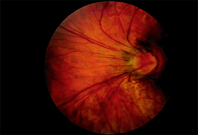
Choroidal folds are lines, grooves or striae predominantly involving the posterior pole of the eye which appear as alternating light and dark lines on fluorescein angiography. They are often arranged in a parallel and horizontal fashion but may be vertical, oblique, or irregular. These folds reflect the undulations of the choroid, retinal pigment epithelium and the overlying neurosensory retina and tend to vary in length and width, rarely extending beyond the equator.
How to tell chorioretinal folds from choroidal folds?
In cases of chorioretinal folds, OCT shows that the retina, RPE and choroid are all folded in a similar manner and maintain their normal relationships and apparently normal thickness, although a certain flattening of the inner retina can be seen along the crests , whereas choroidal folds correspond to undulations of the hyperreflective line corresponding to the RPE as well as the underlying choroid with a flat or nearly flat retinal surface with a variable retinal thickness variable, increasing in correspondence to the valleys and reducing in the crests. The retina, which has a softer structure, adapts itself to the wrinkling to preserve a flat inner surface.
What phase of the venous phase do folds appear?
The diagnosis is made mainly by fluorescein angiography, wherein the folds appear as a series of alternating hyperfluorescent and hypofluorescent streaks starting early in the arteriovenous phase, persisting through the late venous phase, and not leaking in the late films. The hyperfluorescent areas correspond to the peaks of the choroidal folds, and the hypofluorescent areas to the valleys.
Why are choroidal folds seen prominently?
The folds are seen prominently due to their classic alternating hypo and hyperfluorescence. Fluorescein angiography cannot differentiate chorioretinal from choroidal folds, but the optical coherence tomography (OCT) patterns of the two are strikingly different although these are not completely singular entities.
What causes folds in the posterior choroid?
While the term “idiopathic” means the cause of the folds is unknown, previous authors proposed that such eyes may have had prior episodes of insidious or chronic inflammation of the orbit or paranasal sinuses or posterior scleritis, thus leading to permanent thickening of the posterior sclera and flattening of the posterior globe and resulting in relaxation and eventually compression of the posterior choroid, creating folds. In some cases of idiopathic choroidal folds, B-scan ultrasonography has been reported to show flattening of the posterior ocular wall, thought to be due to thickening of the ocular coats.
Is choroidal folds idiopathic?
In the literature, the two largest case series of choroidal folds as well as many other reports identified hyperopia and AMD as the most common associated conditions. A significant number of cases was considered idiopathic although with improved diagnostic testing, the patients formerly referred to as idiopathic actually represented a smaller portion of the total.
Can choroidal folds occur unilaterally?
Choroidal folds can occur unilaterally or bilaterally. Orbital tumors, intraocular tumors, hypotony, scleral buckling, trauma are asscoiated with unilateral folds whereas thyroid disease, systemic medication, choroiditis are more commonly found in bilateral folds. Hyperopia and AMD can be seen in both unilateral and bilateral cases of folds.
ICD-10 Equivalent of 743.5
As of October 2015, ICD-9 codes are no longer used for medical coding. Instead, use this equivalent ICD-10-CM code, which is an exact match to ICD-9 code 743.5:
Historical Information for ICD-9 Code 743.5
Non-Billable means the code is not sufficient justification for admission to an acute care hospital when used a principal diagnosis. Use a child code to capture more detail.
What are retinal folds?
Retinal folds also tend to have less-regular patterns and are often associated with traction in the form of an epiretinal membrane or chorioretinal scar. They may be concentric around a swollen optic disc, in which case they are referred to as Paton's lines.
What are the differences between chorioretinal folds and retinal folds?
The latter are typically finer, have less color, and unlike chorioretinal folds are not seen on fluorescein angiography.
What are the mechanisms that explain the formation of chorioretinal folds?
These theories have included choroidal congestion and an anatomic attachment of Bruch's membrane to the underlying choriocapillaris, 3 change in the tension of Bruch's membrane with secondary involvement of the RPE, 4 and any factor that forces Bruch's membrane to fold. 5 Chorioretinal folds have also been attributed to biomechanical properties in the choroid and sclera and their response to compressive stress. 6
What is the mnemonic for chorioretinal folds?
We have, therefore, found the simple mnemonic, "THIN RPE," quite useful. The mnemonic is easy to remember since chorioretinal folds produce "THIN RPE" at the crest of the fold, thereby producing angiographic hyperfluorescence. The causes, each covered in turn below, are as follows:
Why is it important to recognize chorioretinal folds?
Recognizing chorioretinal folds is important, therefore, not just as part of the complete eye examination, but as an essential part of the diagnostic workup of patients with this distinctive fundus finding.
When were chorioretinal folds first described?
Chorioretinal folds were first described in 1884 by Edward Nettleship in a patient with atrophic papilledema caused by an intracranial mass. 1 Although now known to be uncommon, chorioretinal folds are quite characteristic when observed. Moreover, and perhaps most importantly, chorioretinal folds occur in the setting of a relatively limited number ...
Which imaging technique is used to show chorioretinal folds?
Fluorescein angiogram. Although fluorescein angiography is the preferred method of demonstrating chorioretinal folds, other imaging techniques may be useful as well. Optical coherence tomography, for example, nicely demonstrates the characteristic folds (See Figure 2C).
What are focal folds in dogs?
Focal retinal folds: These are wrinkles in the retinal tissue in one or more areas. They cause small blind spots, but rarely cause visual impairment for the dog. They primarily occur in young dogs with developing retinas, and usually disappear by the age of one year. Young dogs (<1 year of age) with folds only are not at risk of developing dysplasia at a later age.
What is the term for a form of detachment of the retina?
Generalized or complete retinal dysplasia: This form exhibits severe retinal disorganization and is associated with detachment of the retina. Detachment occurs when the retinal sensory tissue separates from the tissue in the back of the eye, thereby causing complete blindness. This is detectable at birth and is permanent.
How long does it take for a puppy to develop retinal dysplasia?
Retinal dysplasia is not a progressive disease, but it will continue to develop until the retinas are mature at around one year of age. To allow the retinas time to mature, the earliest age for a CERF screening for this disease is 6 months, yet closer to one year is recommended by Dr. Kirk Gelatt at which time a definite diagnosis can be established. If you suspect one of your puppies has visual impairment, seek an ophthalmology exam immediately. Signs would include frequently bumping into objects, less activity, and possibly timid or fearful.
What is the layer of sensory tissue that is attached to the tissue in the back of the eye?
The retina is a layer of sensory tissue that is attached to the tissue in the back of the eye. The retina receives light stimuli from the environment and converts it to an electric signal that is sent to the brain via the optic nerve, which lies behind the retina. The brain interprets this signal and vision occurs. Any alterations in the retinal tissue can impair vision. The retinal tissue undergoes changes until the eye is fully developed at the age of one year.
When does retinal dysplasia occur?
It can involve both eyes or one, but the inherited form is almost always bilateral. It can occur anytime during eye development, from pregnancy to one year of age. There are 3 forms of retinal dysplasia:
Can a dog have a rosette?
This may cause some degree of visual impairment and possibly blindness. This can develop at anytime up to one year of age and is usually permanent. In puppies, this can co-exist with folds since folds and dysplasia are difficult to differentiate in the immature retina. As the folds disappear later when the retinas mature, the dysplasia will persist.
Can dogs have retinal folds?
There is no treatment for this disease. The best we can do is to strive to prevent it. The literature recommends that dogs affected with either of the 2 more serious forms of dysplasia (geographic or complete) should not be bred, as well as their parents and their littermates. The genetic relationship between retinal folds and the other 2 forms of dysplasia is unknown at this time. Some ophthalmologists suggest that a mature dog with persistent retinal folds may produce more severe forms of the disease in their offspring, secondary to the dog being a carrier of the gene for the more severe forms of dysplasia. What appear to be folds on exam can sometimes be dysplasia, so a mature dog with folds or dysplasia should not pass a CERF exam. In addition, one may wonder if retinal dysplasia could play a role in the incidence of retinal detachments that occur after bichon cataract surgery. According to Dr. Kirk Gelatt, post-operative detachments are primarily related to ‘cataract maturity and the secondary lens-induced inflammation, rather than retinal dysplasia’.
What is the ICd 10 code for retinal detachment?
361.89 is a legacy non-billable code used to specify a medical diagnosis of other forms of retinal detachment. This code was replaced on September 30, 2015 by its ICD-10 equivalent.
What is the retinal detachment?
It provides the sharp, central vision needed for reading, driving, and seeing fine detail. A retinal detachment lifts or pulls the retina from its normal position. It can occur at any age, but it is more common in people over age 40. It affects men more than women and whites more than African Americans. A retinal detachment is also more likely to occur in people who
Popular Posts:
- 1. icd 10 code for hcoronary artery disease
- 2. icd 10 code for us renal failure
- 3. icd 10 code for abnormal urine findings
- 4. icd 10 code for claustrophobia
- 5. icd 10 code for pvd with gangrene foot
- 6. icd 10 code for seborrheic dermatitis
- 7. icd 9 code for cytotec
- 8. icd code for multifocal intraocular lens
- 9. icd 10 code for cutaneous candidiasis groin
- 10. what is the icd-9 cm code for cauda equina