88.41
What is CPT 75716?
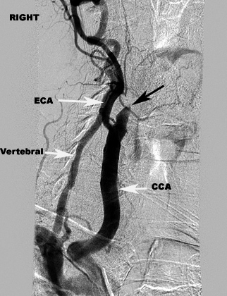
“ Codes 75625 and 75716 are for imaging of the aorta and [bilateral] runoff vessels when the physician images the aorta at one cath position and performs the runoff after moving the catheter to another location ” Zielske says.
What is the CPT code for CT scan with contrast?
cpt codes for ct scans orbit, face & neck 70482 - w/o & w/ contrast maxillofacial/tmj 70486 - w/o contrast 70487 - w/contrast 70488 - w/o & w/ contrast soft tissue neck 70490 - w/o contrast 70491 - w/contrast 70492 - w/o & w/ contrast upper extremity 73200 - w/o contrast 73201 - w/contrast 73202 - w/o & w/ contrast lower extremity 73700 - w/o contrast
What is the CPT code for cardiac monitoring?
CPT code 93228 is the professional component of this service and includes review and interpretation of each 24-hour cardiac surveillance as well as 24-hour availability and response to monitoring events within a course of treatment that includes up to 30 consecutive days of cardiac monitoring.
What is the CPT code for angiography extremity unilateral?
These are very commonly used for Peripheral angiography. There are two CPT® codes for extremity angiogram, code 75710 and 75716. These are mostly used with Non-selective study of abdominal aortogram. The coded 75710 is used for unilateral and 75716 are used for bilateral study of extremity angiogram.
What are ICD-9 procedure codes?
ICD-9-CM is the official system of assigning codes to diagnoses and procedures associated with hospital utilization in the United States. The ICD-9 was used to code and classify mortality data from death certificates until 1999, when use of ICD-10 for mortality coding started.
What is the ICD-9 code for CVA?
ICD-9-CM Diagnosis Code 437.9 : Unspecified cerebrovascular disease.
Is ICD-9 still used in 2020?
Easier comparison of mortality and morbidity data Currently, the U.S. is the only industrialized nation still utilizing ICD-9-CM codes for morbidity data, though we have already transitioned to ICD-10 for mortality.
What is the term for the crosswalk from ICD-9 to ICD-10 or vice versa?
The GEMs are a tool you can use to convert data from ICD-9-CM to ICD-10-CM and ICD-10-PCS and vice versa. The GEMs are also known as crosswalks as they provide important information linking codes of one system with codes in the other system.
What is the ICD-10 for CVA?
I63. 9 - Cerebral infarction, unspecified | ICD-10-CM.
Is a cerebral infarction the same as a stroke?
A cerebral infarction (also known as a stroke) refers to damage to tissues in the brain due to a loss of oxygen to the area. The mention of "arteriosclerotic cerebrovascular disease" refers to arteriosclerosis, or "hardening of the arteries" that supply oxygen-containing blood to the brain.
Why are ICD-9 codes no longer used?
Why the move from ICD-9 codes to ICD-10 codes? The transition for medical providers and all insurance plan payers is a significant one since the 18,000 ICD-9 codes are to be replaced by 140,000 ICD-10 codes. ICD-10 replaces ICD-9 and reflects advances in medicine and medical technology over the past 30 years.
What are the major differences between ICD-9 and ICD-10?
ICD-9 uses mostly numeric codes with only occasional E and V alphanumeric codes. Plus, only three-, four- and five-digit codes are valid. ICD-10 uses entirely alphanumeric codes and has valid codes of up to seven digits.
When did we stop using ICD-9 codes?
No updates have been made to ICD-9 since October 1, 2013, as the code set is no longer being maintained.
What maps ICD-9-CM codes to ICD-10-CM codes?
The GEMs are a tool that can be used to convert data from ICD-9-CM to ICD-10-CM and PCS and vice versa. Mapping from ICD-10-CM and PCS codes back to ICD-9-CM codes is referred to as backward mapping. Mapping from ICD-9-CM codes to ICD-10-CM and PCS codes is referred to as forward mapping.
What type of code may be used when two diagnoses or a diagnosis with a secondary process is present?
Combination Codes: single code used to identify two diagnoses, or a diagnosis with a secondary process or manifestation, or a diagnosis with an associated complication.
What is an ICD-10 crosswalk?
Video: Crosswalking Crosswalking is the mapping of equivalent or near-equivalent codes between two different code sets. The most important type of crosswalking today is between ICD-10-CM and ICD-9-CM.
What is the CPT code for a thoracic angiogram?
This angiogram is very rarely coded. The CPT® code for Thoracic angiogram is 75605 and should be coded only when the document clearly supports for the thoracic angiogram. Most of the times we get confused, whether it is an arch angiogram or thoracic angiogram. So, do check the documentation to code thoracic angiogram CPT® code 75605.
What is the CPT code for spinal angiography?
So, do remember for using this code, always check the tip of the position of catheter. The tip of the catheter should be in the spinal arteries to code this CPT® code 75706.This Angiogram CPT® code could not be used when the spinal arteries are studied by placing the catheter in different position.
What is the CPT code for a visceral artery?
Visceral Arteries include mesenteric artery, splenic artery, hepatic artery, celiac artery etc. The CPT® code used for visceral angiogram is 75726. This CPT® code includes the abdominal aortogram or angiogram 75625. Therefore, do not code CPT® code 75726 and 75725 together. The code description includes word “SELECTIVE” in it. Hence, should be coded only when the tip of the catheter is present in the visceral arteries.
What is CPT code 36221?
The CPT® codes ranging for 36221 - 36228 comprises of the Non-Selective and Selective Catheterization for Cerebral angiogram. These CPT® codes include the supervision and interpretation for cerebral angiogram and hence should not to be coded separately. Therefore, for Cerebral angiogram we should not be worried, just code for the procedure performed and the respective angiogram, as internal carotid, external carotid or cervicocerebral arch angiogram will be included with that procedure code.
What is a CAD angiogram?
Angiograms are performed primarily to diagnose vascular disease throughout the body. It’s common to see the diagnoses in the list below as the pre/post-operative diagnosis for angiography procedures. Pain in chest/angina. Coronary artery/heart disease (CAD) (CHD) Arterio/atherosclerotic heart disease (ASHD) Ischemic heart disease (IHD) ...
What is the diagnostic angiogram?
Diagnostic angiogram is often performed immediately preceding a therapeutic procedure such an angioplasty or thrombectomy and when looking for disease in the heart, angiography is often accompanied by a diagnostic heart cath.
What are the 6th and 7th character of PCS angiography code?
The 6 th and 7 th character of a PCS angiography code are qualifiers which allow additional explanatory information to be communicated by the code. Some qualifiers and their values are specific to certain imaging “types”. For example, the value of “0” indicates a qualifier of “Unenhanced and Enhanced” for the CT and MRI imaging types but indicates “intraoperative” for the fluoroscopy imaging type. This means qualifier values are not necessarily interchangeable, so the PCS table should always be consulted to determine the correct value to assign.
What is the purpose of angiogram?
Angiography is a radiological procedure that uses fluoroscopy, x-ray, CT or MRI to image arteries and veins in relation to vascular obstructions such as atherosclerosis , embolism or thrombus or vascular anomalies.
What is fluoroscopy in angiography?
Fluoroscopy is the most common type of imaging for angiography.
How many characters are needed to report an angiography procedure?
The following are some of the details about what information the values for the 7 characters used to create an ICD-10-PCS angiography code report.
Where do angiography codes come from?
All angiography codes will come from the “Imaging” section of ICD-10-PCS, but the correct code table will vary based on the value of the Body System character.
Overview
Cerebral angiography is a form of angiography which provides images of blood vessels in and around the brain, thereby allowing detection of abnormalities such as arteriovenous malformations and aneurysms. It was pioneered in 1927 by the Portuguese neurologist Egas Moniz at the University of Lisbon, who also helped develop thorotrast for use in the procedure.
Uses
Cerebral angiography is used for diagnosis but may be followed by treatment procedures in the same setting. Cerebral angiography is used to image various intracranial (within the head) or extracranial (outside the head) diseases.
Intracranial diseases are: non-traumatic subarachnoid haemorrhage, non-traumatic intracerebral haemorrhage, intracranial aneurysm, stroke, cerebral vasospasm, cerebral arteriovenous malform…
Technique
Before the procedure, focused history and neurological examination is performed, available imaging, and blood parameters are reviewed. When reviewing imaging, arch anatomy and variants are evaluated to select suitable catheters to assess the vessels. Complete blood count is reviewed to ensure adequate amount of haemoglobin in subject's body, and to rule out the presence of sepsis. Serum creatinine is assessed to rule out renal dysfunction. Meanwhile, prothrombin time is …
Complications
The most common complication is groin haematoma which occurs in 4% of those affected. Neurologic complications such as transient ischemic attack in 2.5% of the cases. There is also the risk of stroke with permanent neurological defect in 0.1% of the cases and may lead to death in 0.06%. Rarely, 0.3 to 1% of the cases experience cortical blindness from 3 minutes to 12 hours after the procedure. It is a condition where those affected experienced loss of vision with normal pupillary …
History
Cerebral angiography was first described by Antonio Caetano de Abreu Freire, Portuguese physician and politician in 1927. He performed this procedure on six patients. Two developed Horner's syndrome due to leaking of contrast material around the carotid artery, one developed temporary aphasia, and another died due to thromboembolism to the anterior circulation of the brain.
External links
• Cerebral Angiography
Popular Posts:
- 1. icd 10 code for abnormal blood count
- 2. icd 10 code for chronotropic incompetence
- 3. icd 9 code for gastric bypass surgery
- 4. icd 9 code for jejumostomy tube status
- 5. icd 10 code for deep tissue injury sacrum
- 6. icd 10 code for right corneal abrasion
- 7. icd 10 code for renal carcinoma'
- 8. icd 10 code for chronic kidney disease unspecified
- 9. icd 10 code for travel
- 10. icd 10 code for rectal avm