What is the ICD 10 code for vasospasm and vasoconstriction?
Cerebral vasospasm and vasoconstriction. I67.84 should not be used for reimbursement purposes as there are multiple codes below it that contain a greater level of detail. The 2019 edition of ICD-10-CM I67.84 became effective on October 1, 2018.
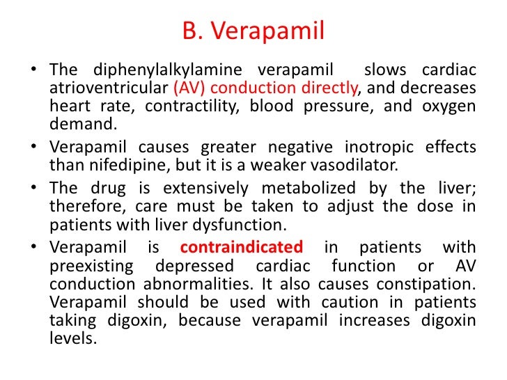
Is intra-arterial administration of verapamil safe for the treatment of cerebral angiospasm?
Endovascular administration of verapamil for treatment of cerebral vasospasm is a safe technique which positively affects the overall recovery of such patients. Keywords: Cerebral angiospasm; Intra-arterial administration of verapamil; Intracranial aneurysm; Subarachnoid hemorrhage; Verapamil.
Are intra-arterial calcium channel blockers safe and efficacious for cerebral vasospasm?
Background: Studies attempting to establish the safety and efficacy of standard and high-dose intra-arterial infusions of calcium channel blockers for treatment of cerebral vasospasm have focused on hemodynamic changes during the angiographic procedure.
Which medications are used to treat vasospasm?
Some commonly infused drugs to treat vasospasm include verapamil, papaverine, milrinone, and nimodipine. The injection of a drug is not separately reported. NEW!

What is the ICD 10 code for vasospasm?
ICD-10 code I67. 84 for Cerebral vasospasm and vasoconstriction is a medical classification as listed by WHO under the range - Diseases of the circulatory system .
What is a cerebral vasospasm?
Cerebral vasospasm is a delayed event after an aneurysmal subarachnoid hemorrhage, with a usual peak from days 7 to 9 after a bleed. It usually affects the large arteries near the ruptured aneurysm.
What medication is used to decrease vasospasm?
Nimodipine has been recommended as first-line medical treatment for preventing post-aSAH cerebral vasospasm. It is usually given orally at a dosage of 60 mg every 4 hours for 21 days after the initial subarachnoid hemorrhage.
What procedure is used to treat cerebral vasospasm?
Treatment for vasospasm can occur through both ICU intervention and endovascular administration of intra-arterial vasodilators and balloon angioplasty. The best outcomes are often attained when these methods are used in conjunction.
Is cerebral vasospasm a stroke?
Subarachnoid Hemorrhage After the hemorrhage, the blood can irritate the brain and cause the vessels in the brain to narrow or go into spasm, limiting blood flow and putting the brain at risk for stroke. This condition is called a cerebral vasospasm.
Is vasospasm the same as vasoconstriction?
A vasospasm is the narrowing of the arteries caused by a persistent contraction of the blood vessels, which is known as vasoconstriction.
Which calcium channel blocker may be used in the treatment of cerebral vasospasm?
Nimodipine. Nimodipine is a dihydropyridine agent that blocks voltage-gated calcium channels and has a dilatory effect on arterial smooth muscle. It is the only FDA-approved agent for vasospasm with a half-life of about 9 h [6].
What is the gold standard for evaluating cerebral vasospasm?
Angiography of the vessels of the brain is the gold standard for the diagnosis of cerebral vasospasm.
What medications cause vasospasm?
SubstancesAngiotensin-Converting Enzyme Inhibitors.Anti-Inflammatory Agents, Non-Steroidal.Antidepressive Agents.Calcium Channel Blockers.Hydroxymethylglutaryl-CoA Reductase Inhibitors.Nitric Oxide Donors.
What causes vasospasm after SAH?
Blockage of the normal CSF circulation can enlarge the ventricles (hydrocephalus), causing confusion, lethargy, and loss of consciousness. Vasospasm is a common complication that may occur 5 to 10 days after SAH (Fig. 2). Irritating blood byproducts cause the walls of an artery to contract and spasm.
How is vasospasm diagnosed after SAH?
Imaging Studies. Various imaging modalities are employed to diagnose cerebral vasospasm after aneurysmal subarachnoid hemorrhage (aSAH). Currently, transcranial Doppler (TCD) is the primary imaging technique used in screening for asymptomatic spasm.
When should nimodipine be given?
Nimodipine comes as a capsule and an oral solution (liquid) to take by mouth or be given through a feeding tube. It is usually taken every 4 hours for 21 days in a row. Treatment with nimodipine should be started as soon as possible, no later than 96 hours after a subarachnoid hemorrhage occurs.
What causes vasospasm in the brain?
Vasospasm occurs when a brain blood vessel narrows, blocking blood flow. It can occur in the two weeks following a subarachnoid hemorrhage or brain aneurysm. You are at greater risk for a cerebral vasospasm if you have had a recent subarachnoid hemorrhage or ruptured brain aneurysm.
Does cerebral vasospasm go away?
Transcranial and Cervical Ultrasound in Stroke Cerebral vasospasm after subarachnoid hemorrhage (SAH) is a preventable and reversible life-threatening condition.
Are Vasospasms serious?
Vasospasm occurs when an artery suddenly narrows and the blood supply is drastically reduced. It most often happens in the brain or in the heart. The results can be serious.
What are the symptoms of vasospasm?
Symptoms of cerebral vasospasm can include:A severe headache.Partial or complete vision loss.Weakness on one side of the body.Convulsions.Confusion and difficulty communicating.Change in consciousness or loss of consciousness.
What is the code for vasospasm?
Vasospasm treatment may require use of a specialized balloon to dilate a vasospastic vessel. Codes 61640-61642 describe this type of balloon dilation; however, when associated with and performed at the same session as the infusion therapy, the balloon dilation codes are bundled into the same territory.
What is the code for a CNS embolization?
Embolizations of the central nervous system (CNS), which includes the brain and spinal cord, is reported with 61624 Transcatheter permanent occlusion or embolization (eg, for tumor destruction, to achieve hemostasis, to occlude a vascular malformation), percutaneous, any method; central nervous system (intracranial, spinal cord) and supervision and interpretation (S&I) code 75894 Transcatheter therapy, embolization, any method, radiological supervision and interpretation. These codes describe coil embolization of a well-defined berry aneurysm or a wide-mouthed aneurysm requiring balloon assistance, and placement of a scaffolding stent (e.g., Neuroform™, Enterprise™, LVIS®, or LVIS® Jr.) or a flow diverter (e.g., Pipeline™ Flex, FRED™). After deployment of a coil, embolic material, a flow diverter, or glue, it is often necessary to determine the results of the embolization using follow-up angiography (75898 Angiography through existing catheter for follow-up study for transcatheter therapy, embolization or infusion, other than for thrombolysis ).#N#Report 75898 for each fully documented follow-up imaging performed during and at the conclusion of a CNS embolization procedure. This code may be submitted more than once per patient encounter for CNS embolizations, with the exception of head and neck (non-CNS) embolizations, which are limited to once per session. Catheter placements or diagnostic imaging (which bundles catheter placements) are separately reported with embolization procedures.#N#Example: A patient with known left middle cerebral artery (MCA) bifurcation aneurysm presents for embolization. Via a right femoral access, a sheath is placed and a guiding catheter is advanced into the left common carotid artery, followed by placement of a microcatheter into the MCA (36217 Selective catheter placement, arterial system; initial third order or more selective thoracic or brachiocephalic branch, within a vascular family ). Guiding angiography delineates the dimensions of the aneurysm. The aneurysm is selected, and a framing coil is placed with follow-up imaging, showing good positioning of the coil without vasospasm or distal vessel embolization (75898). Two more coils are placed to complete embolization (61624, 75894). Completion angiography (75898-59 Distinct procedural service) confirms complete occlusion of the aneurysm without complication.#N#Spinal Embolization#N#Percutaneous transcatheter spinal cord interventions (also CNS) are used primarily to diagnose and treat spinal AVMs. Spinal angiography may be initially performed, followed by embolization. Both are reported at the same session when the imaging is diagnostic in nature. The embolization codes remain 61624 and 75894. The spinal cord is considered to be one surgical site, and is coded as one embolization procedure, even if multiple vessels are embolized.
What are the changes in CPT for 2016?
For 2016, the biggest CPT® coding changes affecting interventional radiology occur within the subspecialties of urinary, biliary, and neurologic intervention. In March, we covered urinary intervention and in April we covered percutaneous biliary interventional coding. This month, we’ll finish our series by focusing on transcatheter neuro-interventions and describing three new codes for 2016.#N#Neuro-interventional procedures are focused on the percutaneous treatment of the central nervous system (brain and spinal cord), the head and neck region, and the spine. Highly trained subspecialty physicians — who focus on transcatheter techniques to diagnose and treat pathology in these complex locations — perform the procedures. Abnormalities treated include aneurysms, arterial-venous malformations (AVMs), vasospasm, stroke, stenoses, and tumors.

Popular Posts:
- 1. icd 10 code for sugar in urine
- 2. icd 10 code for cardiac ablation diagnosis
- 3. icd 10 code for refractory epilepsy
- 4. icd 9 code for new onset type 2 diabetes
- 5. icd code for turtle neck safety collars
- 6. icd 10 code for nondisplaced left subcapital femoral neck fracture
- 7. what is the icd 10 code for debridement
- 8. icd 10 code for autosomal dominant polycystic kidney disease
- 9. icd 10 cm code for pain left ankle
- 10. icd 10 code for massive umbilical hemorrhage of newborn