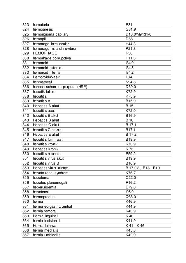
ICD-10 code D18. 0 for Hemangioma is a medical classification as listed by WHO under the range - Neoplasms .
How to code medical diagnosis?
- Point out the tests that were already performed to show the reason for the problem.
- Explain how these evaluations confirmed your diagnosis and show conclusive evidence.
- Use factual information, such as test result quotes, to back up your identification of the patient's issue.
What are DSM diagnosis codes?
Mental retardation
- 317 Mild mental retardation
- 318.0 Moderate mental retardation
- 318.1 Severe mental retardation
- 318.2 Profound mental retardation
- 319 Mental retardation; severity unspecified
What do these diagnosis codes mean?
The CPT code describes what was done to the patient during the consultation, including diagnostic, laboratory, radiology, and surgical procedures while the ICD code identifies a diagnosis and describes a disease or medical condition. … CPT codes are more complex than ICD codes. What is a procedure code and why is it used?
What does diagnosis code 78079 mean?
ICD-9-CM 780.79 is a billable medical code that can be used to indicate a diagnosis on a reimbursement claim, however, 780.79 should only be used for claims with a date of service on or before September 30, 2015. For claims with a date of service on or after October 1, 2015, use an equivalent ICD-10-CM code (or codes).
What is a hemangioma tumor?
A hemangioma (he-man-jee-O-muh) is a bright red birthmark that shows up at birth or in the first or second week of life. It looks like a rubbery bump and is made up of extra blood vessels in the skin. A hemangioma can occur anywhere on the body, but most commonly appears on the face, scalp, chest or back.
What is ICD-10 code for liver hemangioma?
Hemangioma of intra-abdominal structures D18. 03 is a billable/specific ICD-10-CM code that can be used to indicate a diagnosis for reimbursement purposes. The 2022 edition of ICD-10-CM D18. 03 became effective on October 1, 2021.
What is hemangioma of skin and subcutaneous tissue?
Hemangiomas of the skin can form in the top layer of skin or in the fatty layer underneath, which is called the subcutaneous layer. At first, a hemangioma may appear to be a red birthmark on the skin. Slowly, it will start to protrude upward from the skin. However, hemangiomas are not usually present at birth.
What is the ICD-10-CM code for a cavernous hemangioma in intracranial structures?
02.
What is hemangioma of intra abdominal structures?
They are benign tumours that arise from embryonic remnants of unipotent angioblastic cells [1]. Although hemangiomas may occur anywhere within the abdomen, including the solid organs, hollow viscera, ligaments, and abdominal wall, the liver is the most common site.
What is the ICD-10 code for liver disease?
ICD-10 Code for Liver disease, unspecified- K76. 9- Codify by AAPC.
Is a hemangioma a lesion?
Hemangiomas are usually painless, red to blue colored lesions on the skin, lips, or inside the mouth. They are often soft to the touch. Most often they are flush with the skin or slightly elevated, but sometimes they grow from a stalk. Superficial lesions may bleed or turn into sores, particularly if bumped or injured.
What is the difference between an Angioma and a hemangioma?
Angiomas are benign growths made of blood vessels or lymphatic vessels, whereas hemangiomas are small growths made of blood vessels only. Cherry angiomas are most commonly associated with adults. Hemangiomas can appear in early infancy through childhood.
Are all hemangiomas benign?
Spinal hemangiomas are benign tumors that are most commonly seen in the mid-back (thoracic) and lower back (lumbar). Hemangiomas most often appear in adults between the ages of 30 and 50. They are very common and occur in approximately 10 percent of the world's population. Most cases show no symptoms.
What is a cavernous angioma?
A cavernoma is a cluster of abnormal blood vessels, usually found in the brain and spinal cord. They're sometimes known as cavernous angiomas, cavernous hemangiomas, or cerebral cavernous malformation (CCM). A typical cavernoma looks like a raspberry.
What is the ICD-10 code for Cavernoma?
Q28. 3 - Other malformations of cerebral vessels | ICD-10-CM.
Is cavernous hemangioma cancerous?
A liver hemangioma (he-man-jee-O-muh) is a noncancerous (benign) mass in the liver made up of a tangle of blood vessels. Also known as hepatic hemangiomas or cavernous hemangiomas, these liver masses are common and are estimated to occur in up to 20% of the population.
What is a benign skin lesion?
The majority of cases are congenital. A benign skin lesion consisting of dense, usually elevated masses of dilated blood vessels. A benign tumor of the blood vessels that appears on skin. A benign vascular neoplasm characterized by the formation of capillary-sized or cavernous vascular channels.
Is morphology included in the category and codes?
In a few cases, such as for malignant melanoma and certain neuroendocrine tumors, the morphology (histologic type) is included in the category and codes. Primary malignant neoplasms overlapping site boundaries.

Popular Posts:
- 1. icd 10 code for knee osteoarthritis unspecified
- 2. icd 10 code for c diff enteritis
- 3. icd 10 pcs code for transmetatarsal amputation of left foot
- 4. icd 10 code for fall from bed striking object
- 5. icd 10 dx code for insomnia
- 6. icd 10 code for chronic obstructive uropathy
- 7. icd 10 code for laceration left ankle
- 8. icd-10-cm code for decubitus ulcer sacral stage 2
- 9. icd 9 code for uti symptoms
- 10. icd 10 code for hx of hemorragic cva