D18.03
What is the ICD 10 code for hemangioma?
Hemangioma of other sites. D18.09 is a billable/specific ICD-10-CM code that can be used to indicate a diagnosis for reimbursement purposes. The 2019 edition of ICD-10-CM D18.09 became effective on October 1, 2018. This is the American ICD-10-CM version of D18.09 - other international versions of ICD-10 D18.09 may differ.
What is the ICD 10 code for intracranial hemorrhage?
2018/2019 ICD-10-CM Diagnosis Code D18.02. Hemangioma of intracranial structures. 2016 2017 2018 2019 Billable/Specific Code. D18.02 is a billable/specific ICD-10-CM code that can be used to indicate a diagnosis for reimbursement purposes.
What is intraosseous hemangioma?
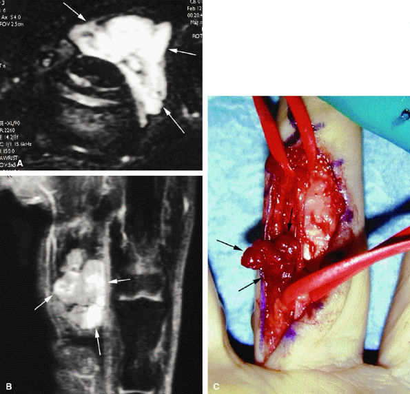
Intraosseous hemangioma is an extremely rare tumor that accounts for 1% or fewer of all osseous tumors. The most common sites of its occurrence are the vertebral column and calvaria. Occurrence in a facial bone is very rare.
What is the ICD 10 code for lymphangioma?
D18 ICD-10-CM Diagnosis Code D18. Hemangioma and lymphangioma, any site 2016 2017 2018 2019 2020 Non-Billable/Non-Specific Code. Type 1 Excludes benign neoplasm of glomus jugulare (D35.6) blue or pigmented nevus (D22.-) nevus NOS (D22.-) vascular nevus (Q82.5) Hemangioma and lymphangioma, any site.
What is the ICD-10 code for hemangioma?
ICD-10 code D18. 0 for Hemangioma is a medical classification as listed by WHO under the range - Neoplasms .
What is the ICD-10 code for a cavernous hemangioma in intracranial structures?
What is the ICD-10-CM code for a cavernous hemangioma in intracranial structures? Rationale: In the ICD-10-CM Alphabetic Index look for Hemangioma/cavernous/intracranial which directs you to D18. 02.
What is hemangioma of intra abdominal structures?
They are benign tumours that arise from embryonic remnants of unipotent angioblastic cells [1]. Although hemangiomas may occur anywhere within the abdomen, including the solid organs, hollow viscera, ligaments, and abdominal wall, the liver is the most common site.
What hemangioma mean?
A hemangioma (hee man jee OH mah) is a common vascular birthmark, made of extra blood vessels in the skin. It is a benign (non-cancerous) growth. The exact cause is not known. Hemangiomas are typically not inherited, but others in the family may also have had them.
What is the ICD-10-CM code for a cavernous hemangioma in intracranial structures D18 00 D18 02 D18 03 q82 5?
Hemangioma of intracranial structures D18. 02 is a billable/specific ICD-10-CM code that can be used to indicate a diagnosis for reimbursement purposes. The 2022 edition of ICD-10-CM D18. 02 became effective on October 1, 2021.
What is a cavernous angioma?
A cavernoma is a cluster of abnormal blood vessels, usually found in the brain and spinal cord. They're sometimes known as cavernous angiomas, cavernous hemangiomas, or cerebral cavernous malformation (CCM). A typical cavernoma looks like a raspberry.
What is intraosseous hemangioma?
Intraosseous hemangioma is a rare bone tumor accounting for 0.7% to 1.0% of all bone tumors. It can occur at all ages but is most common in the fourth and fifth decades of life and has a female preponderance (3:1) [1]. Intraosseous hemangiomas are usually found in the vertebral column and rarely seen in the calvarium.
Is a hemangioma a tumor?
What Is a Hemangioma? Spinal hemangiomas are benign tumors that are most commonly seen in the mid-back (thoracic) and lower back (lumbar). Hemangiomas most often appear in adults between the ages of 30 and 50. They are very common and occur in approximately 10 percent of the world's population.
What is an intra abdominal structure?
Intraperitoneal organs include the stomach, spleen, liver, first and fourth parts of the duodenum, jejunum, ileum, transverse, and sigmoid colon.
What is an atypical hemangioma of the spine?
Rarely, vertebral hemangiomas can exhibit extraosseous expansion with resulting compression of the spinal cord. Such lesions are termed aggressive or atypical vertebral hemangiomas (AVH) and account for less than 1% of spinal hemangiomas.
What is the cause of a hemangioma?
Hemangiomas of the skin develop when there's an abnormal proliferation of blood vessels in one area of the body. Experts aren't sure why blood vessels group together like this, but they believe it's caused by certain proteins produced in the placenta during gestation (the time when you're in the womb).
Do spinal hemangiomas grow?
Benign Spinal Tumors Hemangiomas, Aggressive: A variant of benign hemangiomas, aggressive hemangiomas can increase in size and extend outside the bone into the soft tissue. These may require treatment in some instances.
What causes internal hemangiomas?
An internal hemangioma is a type of noncancerous tumor that forms from the abnormal growth of excess blood vessels. Hemangiomas usually occur on the skin of infants, presenting as a red mark.
What causes hemangiomas to grow?
The female hormone estrogen, which increases during pregnancy, is believed to cause some liver hemangiomas to grow larger. Very rarely, a growing hemangioma can cause signs and symptoms that may require treatment, including pain in the upper right quadrant of the abdomen, abdominal bloating or nausea.
Can a liver hemangioma be misdiagnosed?
When the hepatic hemangioma spontaneously ruptures, it can be easily misdiagnosed because a gastrointestinal perforation is also associated with severe abdominal pain, peritonitis, and shock.
What is capillary hemangioma?
Capillary hemangioma is one of the most common benign orbital tumors of childhood affecting up to 5% of infants under the age of 1 year. It can be superficial, presenting as a red, raised lesion, it can be deep, presenting as a dark blue lesion that may extend into the orbit or may present both of the above components.
Popular Posts:
- 1. icd 10 cm code for no evidence of poisoning or any other signs/symptoms were found
- 2. icd-10 code for left breast wound infection
- 3. icd 10 code for prolonged latent phase of labor
- 4. icd 10 code for pms
- 5. 2016 icd 10 code for adrenal mass
- 6. icd 10 code for vonwillebrand disease
- 7. icd 9 code for carrier of biotinidase def
- 8. icd-10 code for rhabdomyolysis
- 9. icd 10 code for arthritis of bilateral hips
- 10. icd 10 code for posterior scalp hematoma