410.21 - Acute myocardial infarction of inferolateral wall, initial episode of care | ICD-10-CM.
What is isolated lateral wall myocardial infarction?
Continuing Education Activity Isolated lateral wall myocardial infarction (LMI), similar to other acute myocardial infarctions (MI), is caused by acute atherosclerotic plaque rupture with subsequent thrombus formation in the left circumflex (LCx) coronary artery or one of its branches.
What is the ICD 10 version for myocardial infarction of inferior wall?
This is the American ICD-10-CM version of I21.19 - other international versions of ICD-10 I21.19 may differ. Applicable To Acute transmural myocardial infarction of inferior wall
What is the ICD 10 for STEMI of inferior wall?
Short description: STEMI involving oth coronary artery of inferior wall. The 2019 edition of ICD-10-CM I21.19 became effective on October 1, 2018. This is the American ICD-10-CM version of I21.19 - other international versions of ICD-10 I21.19 may differ.
What is the ICD 10 code for lumbar radiculopathy?
I21.29 is a billable/specific ICD-10-CM code that can be used to indicate a diagnosis for reimbursement purposes.
What is the code for Acute myocardial infarction anterior wall?
0 Acute transmural myocardial infarction of anterior wall. Transmural infarction (acute)(of): anterior (wall) NOS.
How do you identify lateral wall MI on ECG?
ECG FindingsST-Elevated LMI: ST elevation in lead I, aVL, V5, and V6; Reciprocal ST depression in inferior lead III and aVF.High lateral STEMI: High lateral STEMI can present as ST-elevation involving lead I and aVL. ... Old LMI presents with deep and broad Q waves I leads I and aVL.More items...•
What is the ICD-10 code for STEMI of inferior wall?
ICD-10-CM Code for ST elevation (STEMI) myocardial infarction of inferior wall I21. 1.
What is anterolateral wall MI?
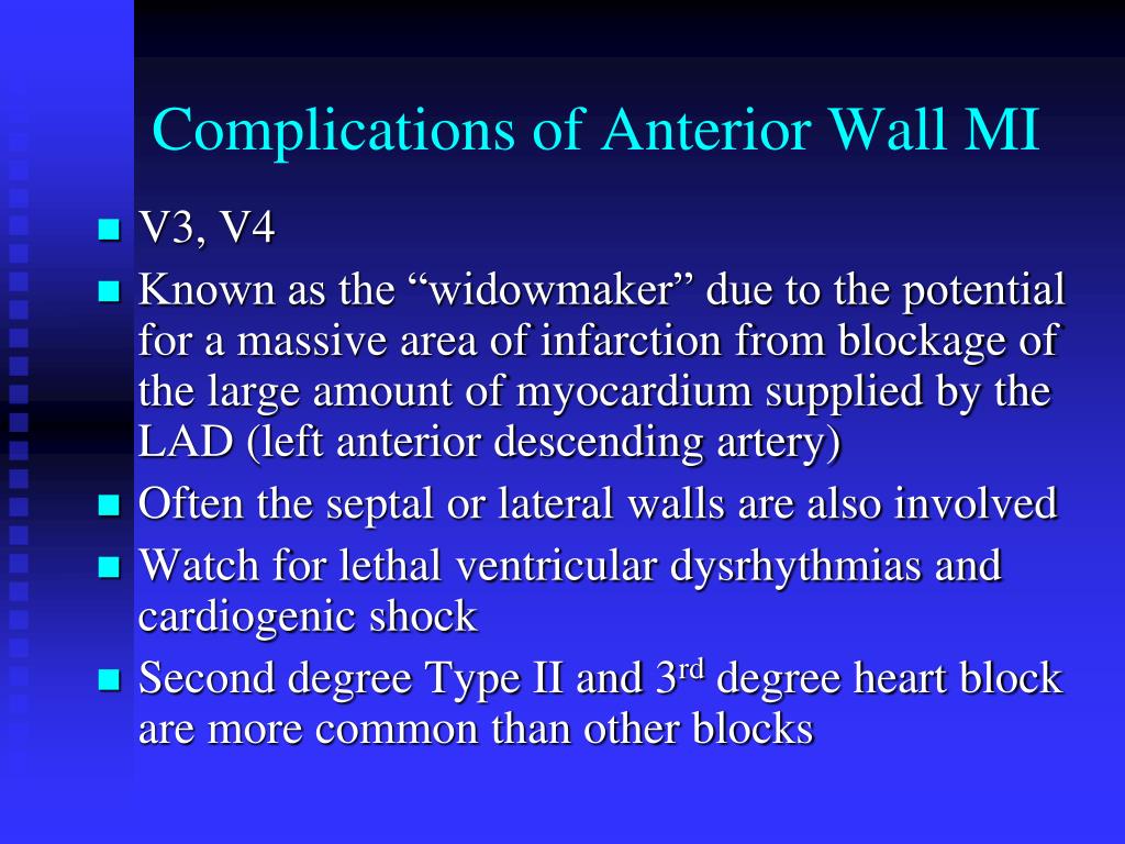
Myocardial infarction in which the anterior wall of the heart is involved. Anterior wall myocardial infarction is often caused by occlusion of the left anterior descending coronary artery. It can be categorized as anteroseptal or anterolateral wall myocardial infarction. [
What is the lateral wall of the heart?
The lateral wall is generally considered to include the wall of the right atrium from the ostia of the superior and inferior vena cava anteriorly to the ostium of the right appendage or auricle.
What is a lateral MI?
A lateral myocardial infarction (MI) is a heart attack or cessation of blood flow to the heart muscle that involves the inferior side of the heart. Inferior MI results from the total occlusion of the left circumflex artery. Lateral MI is characterized by ST elevation on the electrocardiogram (EKG) in leads I and aVL.
What is the ICD-10 code for inferior infarct?
Subsequent ST elevation (STEMI) myocardial infarction of inferior wall. I22. 1 is a billable/specific ICD-10-CM code that can be used to indicate a diagnosis for reimbursement purposes.
What is inferior stemi?
An inferior wall MI — also known as IWMI, or inferior MI, or inferior ST segment elevation MI, or inferior STEMI — occurs when inferior myocardial tissue supplied by the right coronary artery, or RCA, is injured due to thrombosis of that vessel.
What is inferior infarct?
Inferior wall myocardial infarction (MI) occurs from a coronary artery occlusion with resultant decreased perfusion to that region of the myocardium. Unless there is timely treatment, this results in myocardial ischemia followed by infarction.
What is an anterolateral infarction?
Anterolateral infarcts result from the occlusion of the left main coronary artery, and changes appear in leads V5, V6, I, aVL, and sometimes V4. A true anterior infarct doesn't involve the septum or the lateral wall and causes abnormal Q waves or ST-segment elevation in leads V2 through V4.
Where is the lateral wall of the left ventricle?
The free wall of the left ventricle is an area of the ventricular wall which is not in contact with the interventricular septum or apex. It is comprised of the arc of the left atrioventricular groove from the region of continuity between the aortic and mitral valves, to the area lateral of the ventricular septum [8].
What does lateral wall ischemia mean?
It means that blood supply to heart muscle is reduced.Do you have chest pain.
What would be expected when evaluating an ECG for lateral infarction?
There is also subtle ST elevation in the high lateral leads (I and aVL); this may be easily missed. However, the presence of reciprocal ST depression in the inferior leads (III and aVF) makes the lateral ST elevation more obvious. This ECG represents the early stages of a large anterolateral infarction.
What does an MI look like on an ECG?
In the first hours and days after the onset of a myocardial infarction, several changes can be observed on the ECG. First, large peaked T waves (or hyperacute T waves), then ST elevation, then negative T waves and finally pathologic Q waves develop.
What does V1 V2 V3 mean in ECG?
The areas represented on the ECG are summarized below: V1, V2 = RV. V3, V4 = septum. V5, V6 = L side of the heart. Lead I = L side of the heart.
What does ST elevation in lateral leads mean?
ST elevation refers to a finding on an electrocardiogram wherein the trace in the ST segment is abnormally high above the baseline.
What is the code for myocardial infarction?
Codes. I21 Acute myocardial infarction.
What is a myocardial disorder?
A disorder characterized by gross necrosis of the myocardium; this is due to an interruption of blood supply to the area.
What is the ICd 10 code for acute myocardial infarction?
Acute myocardial infarction, unspecified 1 I21.9 is a billable/specific ICD-10-CM code that can be used to indicate a diagnosis for reimbursement purposes. 2 The 2021 edition of ICD-10-CM I21.9 became effective on October 1, 2020. 3 This is the American ICD-10-CM version of I21.9 - other international versions of ICD-10 I21.9 may differ.
When will ICD-10-CM I21.9 be released?
The 2022 edition of ICD-10-CM I21.9 became effective on October 1, 2021.
What is the lateral wall of myocardial infarction?
Isolated lateral wall myocardial infarction (LMI), similar to other acute myocardial infarctions (MI), is caused by acute atherosclerotic plaque rupture with subsequent thrombus formation in the left circumflex (LCx) coronary artery or one of its branches. More commonly the left anterior descending (LAD) coronary artery is involved in the ensuing anterolateral MI. In patients with recent drug-eluting stents, medication non-compliance can cause stent restenosis resulting in acute MI. This activity reviews the medical team's role in evaluating acute lateral wall myocardial infarction and in treating this condition.
What is the left ventricular lateral wall?
The left ventricular lateral wall is supplied by branches of the left anterior descending artery (LAD) and left circumflex artery (LCx). Lateral and posterior walls together form the left ventricular free wall which is a common site for free-wall rupture (FWR) post-MI. Isolated lateral wall involvement is sporadic and is usually seen as part of multi-territorial infarction such as anterolateral, posterolateral, and inferolateral MI. Occlusion of the obtuse marginal branch of the LCx or diagonal branch of LAD can cause isolated lateral myocardial infarction (LMI).
What causes isolated LMI?
Isolated LMI, similar to other acute MI, is caused by acute atherosclerotic plaque rupture with subsequent thrombus formation in LCx or one of its branches . More commonly, LAD is involved in the ensuing anterolateral MI. In patients with recent drug-eluting stents, medication non-compliance can cause stent restenosis resulting in acute MI. On rare occasions, patients can present with myocardial infarction with non-obstructive coronary arteries (MINOCA). The prevalence of MINOCA is estimated at around 6% based on a meta-analysis done in 2015.[2] Some of the causes of MINOCA include:
What waves does LMI present?
Old LMI presents with deep and broad Q waves I leads I and aVL
What is high lateral STEMI?
High lateral STEMI: High lateral STEMI can present as ST-elevation involving lead I and aVL. Subtle ST elevation in V5, V6 and reciprocal changes in lead III and avF may be present. This is usually caused by occlusion of the first diagonal branch of LAD and is sometimes referred to as the South African flag sign.
What should be done for acute LMI?
Patients suspected of having acute LMI should have a prompt evaluation with an electrocardiogram (ECG) and measurements of serial cardiac troponins.[8] Recognizing distinct ST-T involvement patterns can aid in early diagnosis of MI.
What are the symptoms of MI?
Some of the common presenting symptoms include left-sided chest pain radiating to arm/neck, shortness of breath, nausea, vomiting, palpitation, diaphoresis, and fatigue. People with a history of previous MI can relate current symptoms to previous episodes and tend to seek care sooner. Silent MI refers to a group of patients presenting with no acute signs or symptoms. The prevalence of silent MI is reported as high as 23%. [6]
What is the ICD-10 code for myocardial infarction type 2?
I21.A1 is a valid billable ICD-10 diagnosis code for Myocardial infarction type 2 . It is found in the 2021 version of the ICD-10 Clinical Modification (CM) and can be used in all HIPAA-covered transactions from Oct 01, 2020 - Sep 30, 2021 .
Do you include decimal points in ICD-10?
DO NOT include the decimal point when electronically filing claims as it may be rejected. Some clearinghouses may remove it for you but to avoid having a rejected claim due to an invalid ICD-10 code, do not include the decimal point when submitting claims electronically. See also: Infarct, infarction.

Popular Posts:
- 1. 2017 icd 10 code for subglottic aspiration
- 2. icd 10 pcs code for bilateral cardiac catheterization and stenting
- 3. icd 10 code for apc-3 pelvic ring injury
- 4. what is the icd 10-cm code for mild non-proliferative diabetic retinopathy with macular edema
- 5. icd 9 code for schatzki ring
- 6. icd 10 code for sensitive to light
- 7. icd-10 code for coughing
- 8. icd 10 code for atm mutation
- 9. icd 10 code for current use of insulin
- 10. icd 10 code for subtherapeutic inr