What are the new ICD 10 codes?
The new codes are for describing the infusion of tixagevimab and cilgavimab monoclonal antibody (code XW023X7), and the infusion of other new technology monoclonal antibody (code XW023Y7).
What is the ICD 10 code for diastolic dysfunction?
Unspecified diastolic (congestive) heart failure
- Index to Diseases and Injuries. The Index to Diseases and Injuries is an alphabetical listing of medical terms, with each term mapped to one or more ICD-10 code (s).
- Approximate Synonyms
- Convert I50.30 to ICD-9 Code
What are ICD 10 codes?
Why ICD-10 codes are important
- The ICD-10 code system offers accurate and up-to-date procedure codes to improve health care cost and ensure fair reimbursement policies. ...
- ICD-10-CM has been adopted internationally to facilitate implementation of quality health care as well as its comparison on a global scale.
- Compared to the previous version (i.e. ...
What is the ICD 10 diagnosis code for?
The ICD-10-CM is a catalog of diagnosis codes used by medical professionals for medical coding and reporting in health care settings. The Centers for Medicare and Medicaid Services (CMS) maintain the catalog in the U.S. releasing yearly updates.

Is VSD the same as a heart murmur?
A physical exam is one of the most common ways for a doctor to discover a VSD. That's because a VSD — when it's large enough —causes a sound called a heart murmur that your doctor can hear when listening to your heart with a stethoscope. It's even possible to estimate the size of the defect from the sound of a murmur.
What is a VSD in the heart?
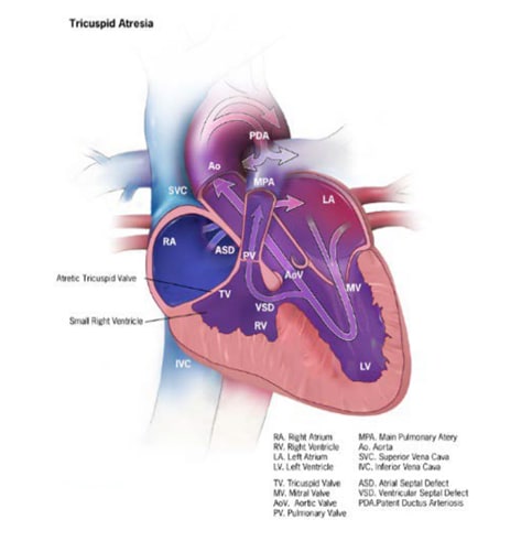
A ventricular septal defect (pronounced ven·tric·u·lar sep·tal de·fect) (VSD) is a birth defect of the heart in which there is a hole in the wall (septum) that separates the two lower chambers (ventricles) of the heart.
What diagnosis is VSD?
About Ventricular Septal Defect Like an atrial septal defect (ASD), a VSD involves a hole, but instead of being located in the wall between the upper atrial chambers of the heart, a VSD occurs in the wall between lower ventricular chambers of the heart. The ventricles are the stronger pumping chambers of the heart.
Is VSD congenital or ASD?
An atrial septal defect (ASD) is a hole in the wall between the heart's two upper chambers. ASD is a congenital condition, which means it is present at birth. A ventricular septal defect (VSD) is a hole in the wall between the two lower chambers. In children, a VSD is usually congenital.
What are the 4 types of VSD?
There are four basic types of VSD:Membranous VSD. An opening in a particular area of the upper section of the ventricular septum (an area called the membranous septum), near the valves. ... Muscular VSD. ... Atrioventricular canal type VSD. ... Conal septal VSD.
What is the most common type of VSD?
Type 2: (membranous) This VSD is, by far the most common type, accounting for 80% of all defects. It is located in the membranous septum inferior to the crista supraventricularis. It often involves the muscular septum when it is commonly known as perimembranous.
What is a small VSD?
A ventricular septal defect (VSD) is a hole in the ventricular septum, the lower wall of the heart separating the right and left ventricles. A VSD is a congenital heart defect, in other words, a birth defect of the heart.
What is the cause of VSD?
The most common cause of a VSD is a congenital heart defect, which is a defect from birth. Some people are born with holes already present in their heart. They may cause no symptoms and take years to diagnose. A rare cause of a VSD is severe blunt trauma to the chest.
What is VSD surgery?
Ventricular septal defect (VSD) surgery is a type of heart surgery. It's done to correct a hole between the left and right ventricles of the heart.
Which is more common ASD or VSD?
Congenital heart defects affect slightly less than 1% of liveborn infants. Two defects,ventricular septal defect (VSD) and atrial septal defect (ASD), account for about 30% of congenital heart disease: VSD for 20% and ASD for 10%.
What is the most common congenital heart defect in adults?
Ventricular septal defect (VSD) (see Figures 2 and 3) is the most common congenital heart defect. Symptoms depend on the size of the defect and the age of the patient.
What is hole in heart called?
An atrial septal defect (pronounced EY-tree-uhl SEP-tuhl DEE-fekt) is a birth defect of the heart in which there is a hole in the wall (septum) that divides the upper chambers (atria) of the heart.
What is a VSD?
A ventricular septal defect (VSD) is a defect in the ventricular septum, the wall dividing the left and right ventricles of the heart. "Illustration showing various forms of a ventricular septal defects. 1. Conoventricular, malaligned 2.
What is the ICD code for ventricular septal defect?
Q21.0 is a billable ICD code used to specify a diagnosis of ventricular septal defect. A 'billable code' is detailed enough to be used to specify a medical diagnosis.
What is billable code?
Billable codes are sufficient justification for admission to an acute care hospital when used a principal diagnosis. The Center for Medicare & Medicaid Services (CMS) requires medical coders to indicate whether or not a condition was present at the time of admission, in order to properly assign MS-DRG codes.
What does "undetermined" mean in medical terms?
Clinically undetermined. Provider unable to clinically determine whether the condition was present at the time of inpatient admission.
When will the ICd 10 Z87.74 be released?
The 2022 edition of ICD-10-CM Z87.74 became effective on October 1, 2021.
What is a Z77-Z99?
Z77-Z99 Persons with potential health hazards related to family and personal history and certain conditions influencing health status
When will the ICd 10 Z82.79 be released?
The 2022 edition of ICD-10-CM Z82.79 became effective on October 1, 2021.
What is a Z77-Z99?
Z77-Z99 Persons with potential health hazards related to family and personal history and certain conditions influencing health status
When will the 2022 ICd-10-CM Q25.0 be released?
The 2022 edition of ICD-10-CM Q25.0 became effective on October 1, 2021.
What is abnormal persistence of an open lumen in the ductus arteriosus after birth?
Abnormal persistence of an open lumen in the ductus arteriosus after birth, the direction of flow being from the aorta to the pulmonary artery, resulting in recirculation of arterial blood through the lungs. Present On Admission. POA Help.
What is the descending aorta?
A congenital heart defect characterized by the persistent opening of fetal ductus arteriosus that connect s the pulmonar y artery to the descending aorta (aorta, descending) allowing unoxygenated blood to bypass the lung and flow to the placenta. Normally, the ductus is closed shortly after birth.
What is the ICd 10 code for ASD?
This is a rare type of ASD and accounts for less than 1 percent cases. Relevant ICD-10-CM codes for ASD are: Q21.1 Atrial septal defect – Alternative wording ...
What is the most common type of ASD?
There are four major types of ASD: Ostium secundum ASD results from incomplete adhesion between the flap valve associated with the foramen ovale and the septum secundum after birth. This is the most common type, accounting for 75 percent of all ASD cases.
What is the most commonly recognized congenital cardiac anomaly presenting in adulthood?
Print Post. Atrial septal defect (ASD) is the most commonly recognized congenital cardiac anomaly presenting in adulthood. An ASD is a defect in the interatrial septum that allows pulmonary venous return from the left atrium to pass directly to the right atrium.
What type of defect must be documented in an AMI?
Documentation must state the exact type of defect the patient has (e.g., type I, type II), and if the condition is congenital or acquired. The contributing factors will indicate the presence of the condition in the setting of an AMI.
What causes Ostium primum ASD?
Ostium primum ASD are caused by incomplete fusion of septum primum with the endocardial cushion. This is the second most common type, accounting for 15-20 percent of cases. Sinus venosus ASD is an abnormal fusion between the embryologic sinus venosus and the atrium. In most cases, the defect lies superior in the atrial septum near the entry ...

Popular Posts:
- 1. icd 10 cm code for other conjunctivitis
- 2. icd 10 code for right cva pain
- 3. icd 10 cm code for transient change in mental status
- 4. icd 10 diagnosis code for history of prostate cancer
- 5. icd 10 code for uric acid crystals
- 6. icd 10 code for alzheimer's with dementia
- 7. 2019 icd 10 code for sudden right hearing loss
- 8. icd 10 pcs code for transvaginal hysterectomy
- 9. icd 10 code for precordial chest pain
- 10. icd code for rt knee chondral