Full Answer
What is the ICD 10 code for uterine fibroids?
Other specified noninflammatory disorders of uterus. N85.8 is a billable/specific ICD-10-CM code that can be used to indicate a diagnosis for reimbursement purposes. The 2020 edition of ICD-10-CM N85.8 became effective on October 1, 2019. This is the American ICD-10-CM version of N85.8 - other international versions of ICD-10 N85.8 may differ.
What is the ICD 10 code for pelvis deformity?
2018/2019 ICD-10-CM Diagnosis Code M95.5. Acquired deformity of pelvis. M95.5 is a billable/specific ICD-10-CM code that can be used to indicate a diagnosis for reimbursement purposes.
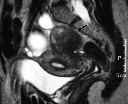
What is the ICD 10 code for calcification of the uterus?
Diagnosis Index entries containing back-references to N85.8: Atresia, atretic uterus Q51.818 ICD-10-CM Diagnosis Code Q51.818 Atrophy, atrophic (of) endometrium (senile) N85.8 Boggy uterus N85.8 Calcification uterus N85.8 Cicatrix (adherent) (contracted) (painful) (vicious) L90.5 - see also Scar ICD-10-CM Diagnosis Code L90.5
What is the ICD 10 code for urethral fibrillation?
D25.9 is a billable/specific ICD-10-CM code that can be used to indicate a diagnosis for reimbursement purposes. The 2021 edition of ICD-10-CM D25.9 became effective on October 1, 2020. This is the American ICD-10-CM version of D25.9 - other international versions of ICD-10 D25.9 may differ.

What is the ICD-10 code for calcifications in the pelvis?
ICD-10 code: M61. 95 Calcification and ossification of muscle, unspecified Pelvic region and thigh.
What is the ICD-10 code for calcified uterine fibroid?
Leiomyoma of uterus, unspecified The 2022 edition of ICD-10-CM D25. 9 became effective on October 1, 2021.
What is the ICD-10 code for calcification?
ICD-10-CM Code for Calcification and ossification of muscle, unspecified M61. 9.
What is the ICD-10 code for fibroids?
9.
What's the difference between a fibroid and a tumor?
A uterine fibroid is the most common benign (not cancerous) tumor of a woman's uterus (womb). Fibroids are tumors of the smooth muscle found in the wall of the uterus. They can develop within the uterine wall itself or attach to it. They may grow as a single tumor or in clusters.
What is the difference between a leiomyoma and a fibroid?
Uterine fibroids are noncancerous growths of the uterus that often appear during childbearing years. Also called leiomyomas (lie-o-my-O-muhs) or myomas, uterine fibroids aren't associated with an increased risk of uterine cancer and almost never develop into cancer.
What is extensive vascular calcification?
Vascular calcifications are mineral deposits on the walls of your arteries and veins. These mineral deposits sometimes stick to fatty deposits, or plaques, that are already built up on the walls of a blood vessel. Vascular calcifications are common but potentially serious.
What is calcified bone?
Calcium is one of the most abundant minerals in the body. It is present in the bones, teeth, and bloodstream. If calcium deposits form, the medical names for this is “calcification.” Calcification can occur with age, but it can also be linked with infections, injuries, and cancer.
What causes soft tissue calcification?
Soft tissue calcification can be caused by secondary tumoural calcinosis from renal insufficiency, or collagen vascular diseases and by vascular calcifications, either arterial or venous (phlebolith).
What does pedunculated fibroid mean?
Pedunculated fibroids are benign (noncancerous) growths in the uterus. These fibroids are attached to the uterine wall by a stalk-like growth called a peduncle. The main difference between pedunculated fibroids and other fibroids is the peduncle. These fibroids can grow both inside and outside the uterus.
What is an intramural fibroid?
An intramural fibroid is a noncancerous tumor that grows between the muscles of the uterus. There are several types of intramural fibroids: anterior intramural fibroid, located in the front of the uterus. posterior intramural fibroid, located in the back of the uterus.
What is the ICD-10 code for pelvic pain?
ICD-10 code R10. 2 for Pelvic and perineal pain is a medical classification as listed by WHO under the range - Symptoms, signs and abnormal clinical and laboratory findings, not elsewhere classified .
What is calcified fibroid?
What is a calcified fibroid? A calcified fibroid is when a fibroid has reached the final stage of degeneration, or cell death and calcium deposits develop on the remaining fibroid tissue. Fibroids are benign tumors that grow in or on the uterine walls.
What does it mean when a woman's fibroid is calcified?
When a woman’s fibroid (s) calcify, it indicated the end stages of degeneration and she may experience less pain or abnormal periods than during the growth and degeneration periods. When the calcified fibroid is large, it may put pressure on the bladder and bowel causing the need for frequent urination, incontinence issues, constipation, ...
Why are fibroids different from calcified fibroids?
Calcified fibroids are different only because they typically have become larger than the blood supply that was attributed to the growth. Now that the blood supply has been compromised the tumor degenerates and may become smaller.
What is the first step in a fibroids treatment plan?
The first step in any treatment plan should be a thorough diagnostic assessment including a detailed conversation about the symptoms, a pelvic exam, and imaging tests, if necessary. The symptoms common to fibroids can also be caused by other conditions such as uterine polyps, polycystic ovary syndrome, or endometriosis.
Can calcification cause miscarriage?
Calcified fibroids can also cause complications in pregnancy including miscarriage, premature placenta detachment, or breech positioning.
What is a fibrous tumor?
Uterine fibroids are the most common non-cancerous tumors in women of childbearing age . Fibroids are made of muscle cells and other tissues that grow in and around the wall of the uterus, or womb. The cause of fibroids is unknown. Risk factors include being african-american or being overweight.
What is the code for a primary malignant neoplasm?
A primary malignant neoplasm that overlaps two or more contiguous (next to each other) sites should be classified to the subcategory/code .8 ('overlapping lesion'), unless the combination is specifically indexed elsewhere.
Can fibroids cause infertility?
most women with fibroids can get pregnant naturally. For those who cannot, infertility treatments may help. Treatment for uterine fibroids includes medicines that can slow or stop their growth, or surgery.
Popular Posts:
- 1. icd 10 code for family hx of prostate cancer
- 2. icd 10 code for submassive pulmonary embolism
- 3. what is the icd 10 code for acute respiratory insufficiency post op
- 4. icd-10 code for screening pap
- 5. icd 9 code for sarcoidosis with cardiomyopathy
- 6. icd 10 code for thoracic lumbar scoliosis
- 7. icd 9 code for developmental age
- 8. what is the icd 10 code for nasopharyngeal lesion
- 9. icd 10 code for rle wound due to lymphedema
- 10. what is the icd 9 code for hypothyroidism