Other benign neoplasm of skin, unspecified
D23. 9 is a billable/specific ICD-10-CM code that can be used to indicate a diagnosis for reimbursement purposes. The 2022 edition of ICD-10-CM D23. 9 became effective on October 1, 2021.What is the ICD 10 code for benign neoplasm of skin?
Other benign neoplasm of skin, unspecified 2016 2017 2018 2019 2020 2021 Billable/Specific Code D23.9 is a billable/specific ICD-10-CM code that can be used to indicate a diagnosis for reimbursement purposes. The 2021 edition of ICD-10-CM D23.9 became effective on October 1, 2020.
What is the ICD 10 code for excluded nevus?
When a type 2 excludes note appears under a code it is acceptable to use both the code (I78.1) and the excluded code together. blue nevus ( ICD-10-CM Diagnosis Code D22 flammeus nevus ( ICD-10-CM Diagnosis Code Q82.5 hairy nevus ( ICD-10-CM Diagnosis Code D22 melanocytic nevus ( ICD-10-CM Diagnosis Code D22
What is the ICD 10 code for neoplasm of face?
2021 ICD-10-CM Diagnosis Code D23.30: Other benign neoplasm of skin of unspecified part of face. ICD-10-CM Codes. ›. C00-D49 Neoplasms. ›. D10-D36 Benign neoplasms, except benign neuroendocrine tumors. ›. D23- Other benign neoplasms of skin.
What is the ICD 10 code for non-neoplastic neoplasm?
Nevus, non-neoplastic. I78.1 is a billable/specific ICD-10-CM code that can be used to indicate a diagnosis for reimbursement purposes.

What is the ICD 10 code for nevus?
I78.11.
What is melanocytic nevi of unspecified part of face?
Melanocytic nevi are benign neoplasms or hamartomas composed of melanocytes, the pigment-producing cells that constitutively colonize the epidermis.
Is a compound nevus benign or Malignant?
benignCompound Nevi Typically they are light tan to dark brown, dome shaped papules that are 1-10 mm in diameter. Compound Nevi are benign proliferations of melanocytes at the epidermal-dermal junction.
How do you code a dysplastic nevus?
ICD20 Dysplastic Nevi I would use D48. 5 for the dx of dysplastic nevi. Also, if the patient also has a hx of dysplastic nevi, don't forget to include Z86.
Is a melanocytic nevus a mole?
Melanocytic naevi are pigmented moles. The word 'melanocytic' means that they are made up of the cells (melanocytes) which produce the dark pigment (melanin) that gives the skin its colour. Melanocytes clustered together form naevi.
What are benign nevi?
Listen to pronunciation. (NEE-vus) A benign (not cancer) growth on the skin that is formed by a cluster of melanocytes (cells that make a substance called melanin, which gives color to skin and eyes). A nevus is usually dark and may be raised from the skin.
What is compound nevus of skin?
Listen to pronunciation. (KOM-pownd NEE-vus) A type of mole formed by groups of nevus cells found in the epidermis and dermis (the two main layers of tissue that make up the skin).
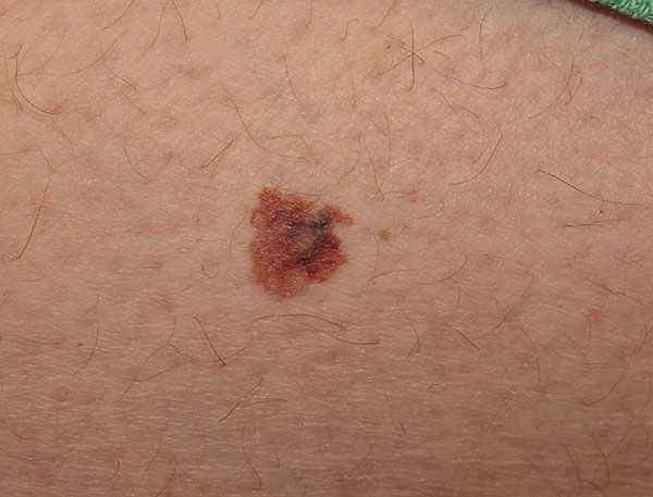
What does a benign nevus look like?
Most people have between 10 and 40. Common nevi are harmless collections of colored cells. They typically appear as small brown, tan, or pink spots. You can be born with moles or develop them later.
What is a benign intradermal nevus?
Intradermal Nevi are benign proliferations of melanocytes in the dermal layer of skin. Intradermal nevi are a common finding in man people, however a change in color, shape, or size should be investigated for malignant transformation.
What is the ICD 10 code for benign nevi?
I78. 1 is a billable/specific ICD-10-CM code that can be used to indicate a diagnosis for reimbursement purposes. The 2022 edition of ICD-10-CM I78.
What is the ICD 10 code for skin lesion?
ICD-10-CM Code for Disorder of the skin and subcutaneous tissue, unspecified L98. 9.
What is non neoplastic nevus?
A abnormal, congenital formation or mark on the skin or neighboring mucosa that does not show neoplastic growth. [
Does melanocytic mean melanoma?
Melanocytes: These are the cells that can become melanoma. They normally make a brown pigment called melanin, which gives the skin its tan or brown color.
What does a melanocytic nevi look like?
They typically appear as small brown, tan, or pink spots. You can be born with moles or develop them later. Moles that you're born with are known as congenital moles. However, most moles develop during childhood and adolescence.
Should melanocytic nevus be removed?
to the editor: In the article on newborn skin, the authors recommend removal of large and giant congenital melanocytic nevi as the current management strategy. In fact, complete nevus removal is impossible for many large nevi and virtually all giant nevi.
What causes melanocytic nevus?
Congenital melanocytic nevi are caused by a change in color (pigment) cells of the skin. The moles happen by chance. CMN is not passed down from the parents. There is no way to prevent your child from being born with moles.
What is the code for a primary malignant neoplasm?
A primary malignant neoplasm that overlaps two or more contiguous (next to each other) sites should be classified to the subcategory/code .8 ('overlapping lesion'), unless the combination is specifically indexed elsewhere.
What chapter is neoplasms classified in?
All neoplasms are classified in this chapter, whether they are functionally active or not. An additional code from Chapter 4 may be used, to identify functional activity associated with any neoplasm. Morphology [Histology] Chapter 2 classifies neoplasms primarily by site (topography), with broad groupings for behavior, malignant, in situ, benign, ...
Can multiple neoplasms be coded?
For multiple neoplasms of the same site that are not contiguous, such as tumors in different quadrants of the same breast, codes for each site should be assigned. Malignant neoplasm of ectopic tissue. Malignant neoplasms of ectopic tissue are to be coded to the site mentioned, e.g., ectopic pancreatic malignant neoplasms are coded to pancreas, ...
What is the code for a primary malignant neoplasm?
A primary malignant neoplasm that overlaps two or more contiguous (next to each other) sites should be classified to the subcategory/code .8 ('overlapping lesion'), unless the combination is specifically indexed elsewhere.
What chapter is neoplasms classified in?
All neoplasms are classified in this chapter, whether they are functionally active or not. An additional code from Chapter 4 may be used, to identify functional activity associated with any neoplasm. Morphology [Histology] Chapter 2 classifies neoplasms primarily by site (topography), with broad groupings for behavior, malignant, in situ, benign, ...
Can multiple neoplasms be coded?
For multiple neoplasms of the same site that are not contiguous, such as tumors in different quadrants of the same breast, codes for each site should be assigned. Malignant neoplasm of ectopic tissue. Malignant neoplasms of ectopic tissue are to be coded to the site mentioned, e.g., ectopic pancreatic malignant neoplasms are coded to pancreas, ...
Popular Posts:
- 1. icd 10 cm 2017 code for cyclosporiasis
- 2. icd 10 code for new injury left knee lateral bucket handle meniscal tear with chondromalacia
- 3. icd 10 code for serveses
- 4. icd 10 code for screening for malignant neoplasm of the ovaries
- 5. icd 10 code for aqueductal stenosis
- 6. icd 10 code for allergy status to nsaids
- 7. icd 10 code for ear rupture
- 8. icd 10 code for history of aaa
- 9. icd 10 code for s/p osteomeitis
- 10. icd-10-pcs code for cardiac catheterization with stent placement